
Get FREE Weekly MRI Content

Traditional teaching methods fall short in two critical areas
MRI physics is abstract. Students read equations and concepts, but they can't visualize how parameters actually affect real images. Theory remains disconnected from practice.
Limited scanner access. Inconsistent quality across sites. No time to experiment. Students watch more than they practice, and they only see routine scans on tight schedules.
"Before Corsmed, there was a big gap between didactic and clinical. Once we implemented Corsmed, that gap was bridged.
Now students learn MRI concepts in a safe space before they enter the higher-stakes clinical setting."

.avif)
Four ways we turn classroom theory into clinical competency
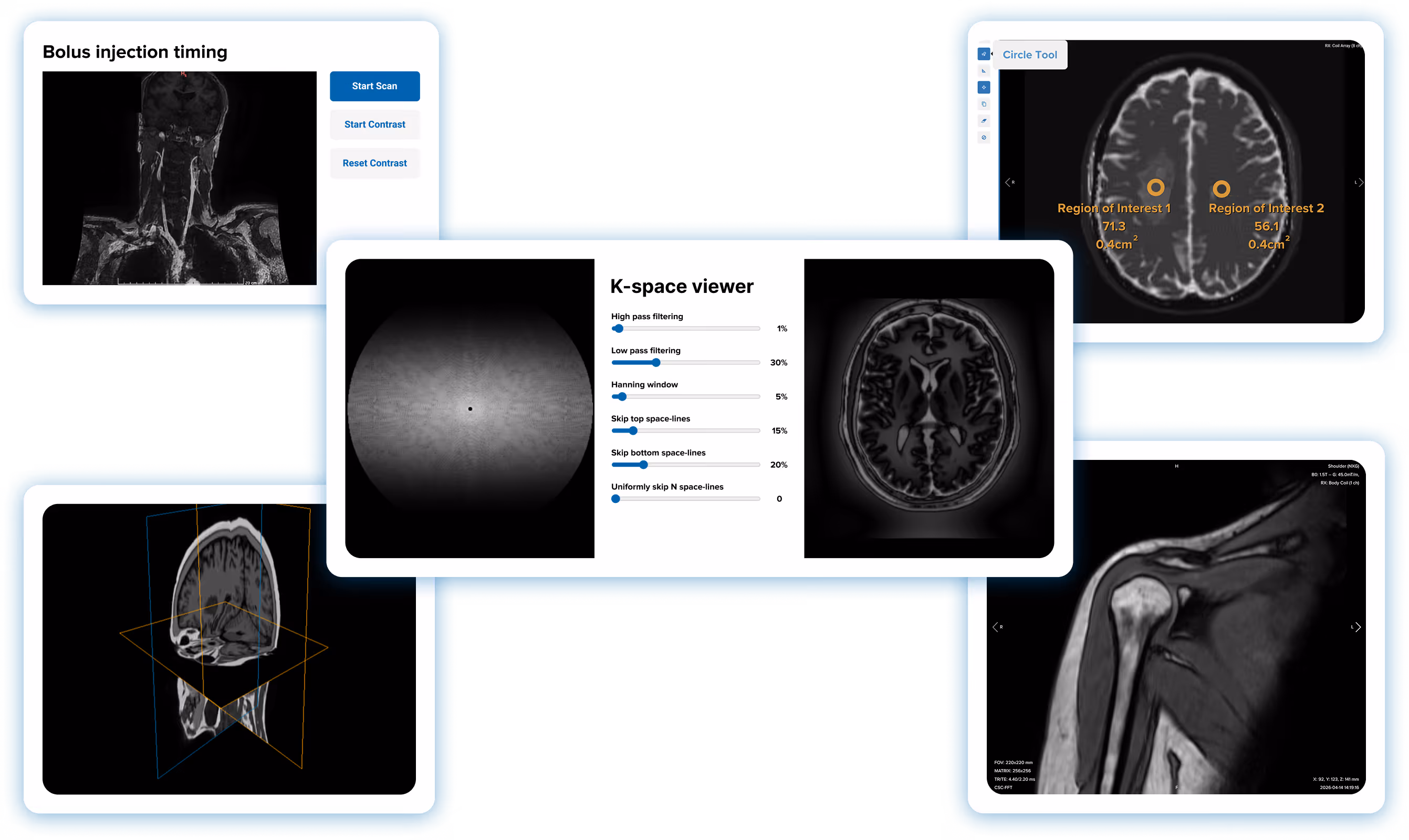
When teaching TR, TE, and flip angles, demonstrate the exact results in real- time. No more imaginary scenarios; students see quantum physics concepts applied in medical practice.
"When teaching MRI physics, I don't have to rely on imaginary scenarios. I can show students the exact result when they apply physics concepts in a medical practice.
This visualization is the main part of Corsmed that helps me teach physics."

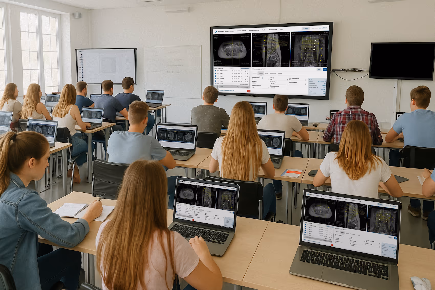
Give students freedom to experiment without patient pressure or scanner time limits. They can practice any anatomy or pathology 24/7 until they master it. Learn by trying, failing, and understanding.
"Corsmed gives students a safe space to learn, free from the pressures of clinical placement.
They can play with the settings, see what changes what, and understand how it affects the image – without worrying about patient care."

Create scanning assignments where students practice real protocols across diverse anatomies and pathologies. They complete hundreds of cases before their first clinical day.
"Using Corsmed, our students complete 500 clinical cases across a wide range of anatomies and pathologies.
When our students began their externships, the imaging centers were amazed at how skilled they were."

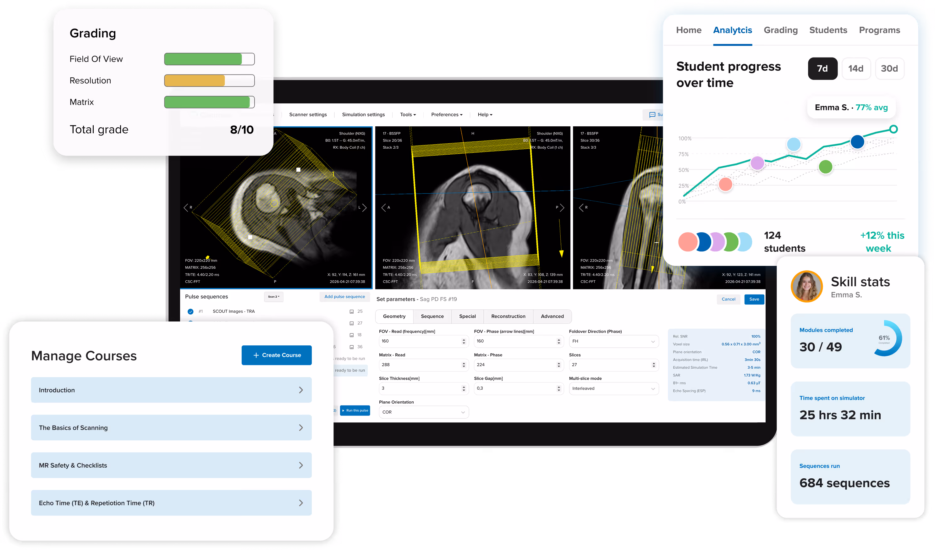
Monitor practical competency with automatic grading, personalized feedback, and detailed analytics. See exactly what parameters students entered, how they prescribed planes, and track their skill development over time.
"Corsmed is an excellent assessment tool because students build protocols and sequences from scratch.
It shows exactly what parameters they've entered and how they've prescribed planes, which helps us test their understanding in many areas."


Colleges and hospitals worldwide use Corsmed to bridge the theory-practice gap

.svg)


Transform your MRI program in 3 straightforward steps
Free expert support and an intuitive platform built for educators
See how Corsmed can transform your MRI program and prepare job-ready graduates.
Fully vendor-neutral. Corsmed teaches universal MRI principles that apply across all major manufacturers: Siemens, GE, Philips, Canon, and others. Students learn MRI physics and concepts, not button positions on a specific console. This is especially useful if you operate scanners from multiple vendors.
Noyal Mathew, MRI Practice educator at University College London Hospitals NHS, especially applauded the vendor-neutral design of Corsmed:
"It's very important that the MRI simulator has a vendor-neutral design. Especially for us at UCLH, where we have many different scanners.
The vendor neutral interface also helps when teaching. I can focus on explaining how the physics work, rather than the placements of buttons.”
Noyal Mathew
MRI Practice Educator, NHS UCLH
You set a scan task. Students then complete it on the simulator and submit their images as the answer. When they submit, you see not just the resulting image stack, but every parameter and slice position they used to produce it.
Grading can be automatic or manual.
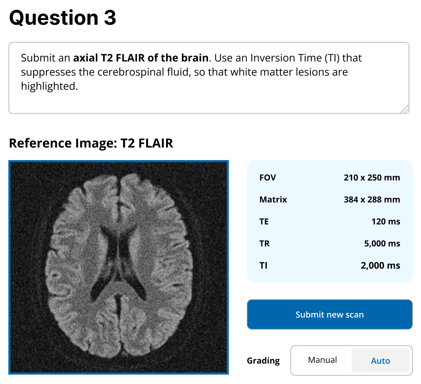
With auto-grading, you perform a reference scan on the simulator yourself. That image becomes the benchmark. Corsmed then automatically compares each student's submission against it and assigns points based on how close they got. A near-perfect result earns full marks. Students get instant feedback without you reviewing every scan individually.
Example of a reference image on an auto-graded scan exercise:
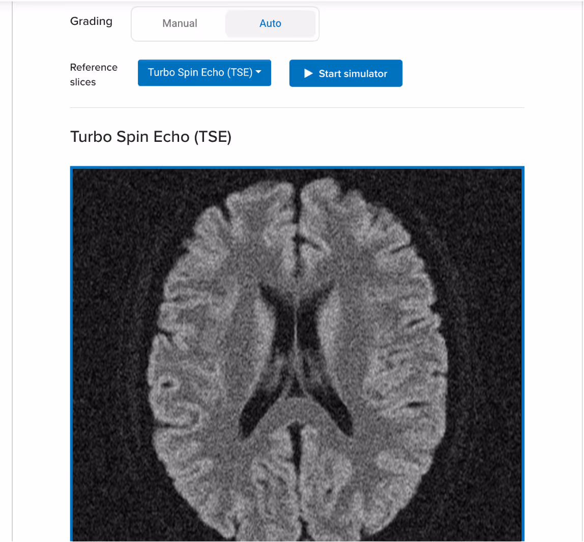
Example of an auto-graded scan:
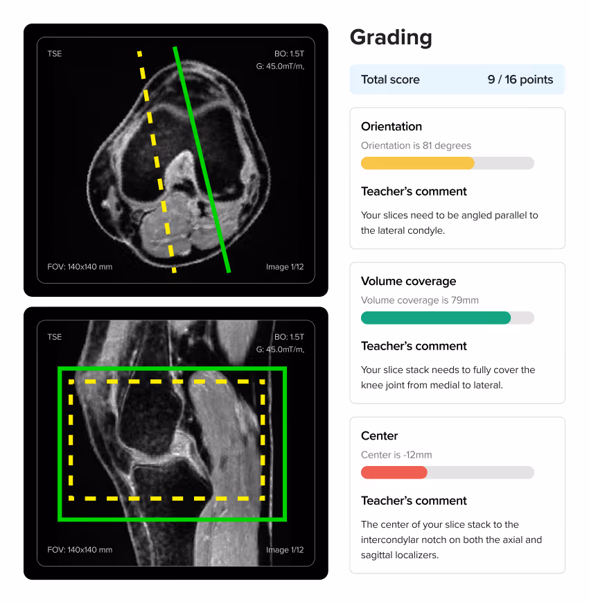
When using auto-grading, you can also specify how the platform should assess students' scans:
Corsmed's auto-grading functionality:

With manual grading, you review each submission yourself and write feedback directly on it, noting exactly what the student got right and what to correct. Students see your annotations alongside their scan.

No, setup is extremely simple. Corsmed works on any computer with an internet connection. There's no software to install, no special hardware to buy, and no IT configuration required. Users just log in and access it through a web browser.
NHS UCLH found that a 1-hour introduction is enough for radiographers to begin practicing independently.
"It's straightforward: press 'Scan' and it scans. Click 'Add protocol' and it adds a protocol.
A 1-hour introduction enables our learners to explore independently. There are good video guides within the 'Help' page too."
Noyal Mathew
MRI Practice Educator, NHS UCLH
Even non-technical users learn to use Corsmed very quickly.
"One of our students wasn't very tech-savvy. She needed IT support just to upload her coursework. But she mastered Corsmed, which shows how user-friendly the simulator is."
Dr. Mahmud Khokhar
MRI Program Director, CNI College
The analytics dashboard gives you a live view of your whole cohort. Module completion, time spent on the simulator, scan count, and grades for every student are visible at a glance.
You can drill into any individual to see exactly where they are in the curriculum and which exercises they've completed. In a self-paced program, this lets you catch who is falling behind before it becomes a problem.


.avif)


.svg)

Give students freedom to experiment without patient pressure or scanner time limits. They can practice any anatomy or pathology 24/7 until they master it. They learn by trying, failing, and understanding.