Train competent MRI technologists faster—without disrupting patient care

The Corsmed MRI simulator enables safe, repeated practice outside the scan room. Teach more effectively, reduce recalled scans, and cut onboarding time by 50%.

Corsmed interfaces showing the MRI simulator, Manage Courses, Grading of scan results, and User Progress analytics.

The MRI simulator that transforms theory into practice. Build courses, monitor student development, and prepare them for clinical success from Day One.

Corsmed interfaces showing the MRI simulator, Manage Courses, Grading of scan results, and User Progress analytics.

Trusted by leading MRI institutions

See the MRI learning platform demo

The challenge when training new MRI technologists

Training new technologists with real scanners falls short at 3 critical points

Calendar icon

Scanners are fully booked with patients

With scanners running 15-20 patients daily, there's no time for hands-on training. New technologists wait for sparse practice opportunities, extending training to 6+ months.

Shield icon

No safe space to make mistakes

With real patients on the table, technologists can't experiment or repeat scans. They need a psychologically safe environment to freely test parameters and build confidence.

Profile icon

Training disrupts patient care

Using clinical scanners for training means taking time away from patients. Educators must also be present during scans to supervise, stealing further time from patient care.

The Corsmed MRI simulator:
Unlimited scanning practice 24/7

Give students freedom to experiment without patient pressure or scanner time limits. They can practice any anatomy or pathology 24/7 until they master it. They learn by trying, failing, and understanding.

"Before Corsmed, there was a big gap between didactic and clinical. Once we implemented Corsmed, that gap was bridged.

Now students learn MRI concepts in a safe space before they enter the higher-stakes clinical setting."

Ayman Darwish, MRI Instructor at NAIT
Ayman Darwish
MRI Instructor, Northern Alberta Institute of Technology
A laptop displaying Corsmed's MRI simulator interface. The screen shows three images of the brain, in axial, sagittal, and coronal planes, with pulse sequence settings being adjusted in the bottom panel.
Explore the MRI simulator
Back arrow icon
Explore the MRI simulator
Back arrow icon

How Corsmed solves your training constraints

We give you what clinical scanners can't; a dedicated training environment that eliminates every barrier between your technologists and confident practice.

1

Practice anytime – without scanner access

Corsmed removes the scanner access bottleneck entirely. Technologists practice scanning anytime, on any laptop, without competing for clinical equipment or affecting a patient appointments.

"The unlimited practice has made our radiographers much more confident in their scanning capabilities.

They understand why, how and which parameters are best to manipulate in order to improve the quality of their scans."

Profile Icon
MRI Manager
The Christie Hospital NHS Foundation Trust
Top image shows a real radiographer operating an MRI scanner while a patient lies inside the bore, with planning images displayed on a clinical console. Bottom image shows a radiographer practicing MRI scanning on the Corsmed MRI simulator using a laptop at home, with simulated planning images visible on the screen.
2

Create psychological safety to experiment

With real patients, technologists can't experiment, can't repeat scans, can't truly learn. Corsmed creates a risk-free learning environment.

Corsmed creates a risk-free learning environment. Students can try any parameters slice planning, create artifacts intentionally, and learn how to optimize images.

"When there's no patient involved, it creates psychological safety.

Technologists can freely test their technical knowledge and skills, and learn without the worry of affecting their patients."

Noyal Mathew – MRI Practice Educator at NHS University College London Hospitals
Noyal Mathew
MRI Practice Educator, University College of London Hospitals NHS Trust
Top image shows a user adjusting scan parameters in the Corsmed MRI simulator. The user selects Turbo Spin Echo, sets TR to 4000 ms, refocusing flip angle to 180 degrees, bandwidth to 781.3 Hz, and lowers NEX/NSA to 1. A blue thought bubble says “What if I lower NEX/NSA?”.
Bottom image shows the resulting elbow MRI, now very grainy, with heavy noise due to the reduced averages. Another blue thought bubble says “Oops, too much noise!”.
3

See parameter effects in real-time

Adjust TR and watch T1 weighting shift. Change bandwidth and see SNR respond. Modify turbo factor and observe the quality-time tradeoff.

"Our technologists felt it was very beneficial to have the simulator and see the effect changes to the parameters made in real time and while they were being taught.

They felt this was more representative of their job."

Profile Icon
MRI Manager
The Christie Hospital NHS Foundation Trust
A laptop displaying Corsmed's MRI simulator interface. The screen shows three images of the brain, in axial, sagittal, and coronal planes, with pulse sequence settings being adjusted in the bottom panel.
4

Assess skills without being present

Create hands-on scan exercises that technologists complete at their own pace, and see exactly what parameters and slice planning they used.

Set up automatic grading, give personal feedback, and track their progress with detailed analytics.

"With the simulator, assessment is easier. Technologists can perform a scan whenever it suits them, and I can review their work later."

Noyal Mathew – MRI Practice Educator at NHS University College London Hospitals
Noyal Mathew
MRI Practice Educator, University College of London Hospitals NHS Trust
Screenshot of a Corsmed cardiac MRI exercise, showing a student's slice planning on two localizer views alongside their scan parameters and an instructor's written feedback.
Screenshot of Corsmed's instructor analytics dashboard, showing student progress over time, with top and underperforming students highlighted by name and average score.

Build custom MRI protocols

Create MRI protocols that match how your team actually scans.

With Corsmed, you can fully customize parameters, workflows, and use cases so training reflects your real clinical setup.

Start from our library of standard protocols, or build your own from scratch. Adapt every detail to fit your organization's needs.

Corsmed interface of the protocol builder where users can create and edit custom MRI scan protocols
"Now, our MRI radiographers gain the same level of competence in just 3-4 months instead of 6. The number of recalls – patients who must redo their scan – has dropped significantly."
Noyal Mathew
MRI Practice Educator, NHS UCLH

Results that matter

When NHS University College London Hospitals and NHS Christie integrated Corsmed into their training programs, they saw measurable improvements in training speed, technologist confidence, and patient care.

2x

Faster training

Technologists reach full competence in 3-4 months instead of 6, through structured practice on the simulator

3x

More practice time

Technologists get triple the hands-on scanning experience without using a single minute of clinical scanner time

Arrow pointing down

Fewer patient recalls

Better parameter understanding and troubleshooting skills reduce repeat scans and patient delays

The complete learning management system built around the simulator

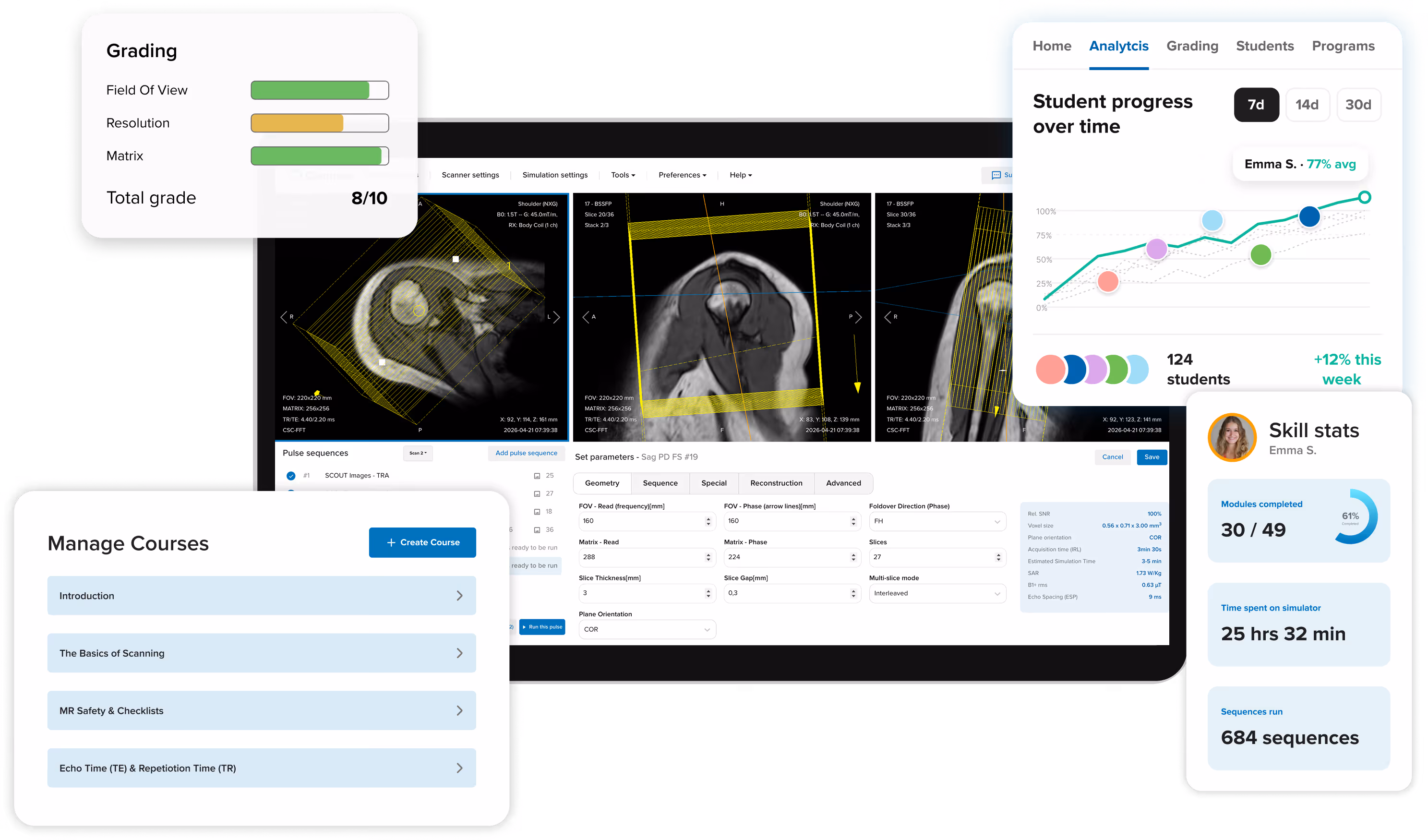
Corsmed is not just a simulator. It's a complete training platform with all the features you need to teach and train MRI effectively: course creation, scan exercises, grading, progress tracking, user management and skill certificates.

Screenshot of Corsmed's training platform showing the MRI simulator at the center of a laptop screen, surrounded by all the features you need to teach effectively, including course modules, scan exercises, automated grading, student analytics, user achievements page, and user access management.
Explore the LMS
Back arrow icon

Tools that help your learners master every part of MRI

In addition to the core simulator, Corsmed offers five additionals tools that help your learners master every aspect of MRI scanning: timing contrast injections, grasping k-space, viewing anatomy in 3D, post-processing, and image review.

A collage of screenshots from Corsmed's MRI training platform showing five imaging tools placed around the MRI simulator: 1) the k-space viewer, 2) the bolus injection timing tool, 3) the 3D viewer showing a rotatable brain model, 4) the post-processing tool with a circular ROIs drawn on a brain scan, and 5) the DICOM viewer showing a shoulder scan
Explore the imaging tools
Back arrow icon

What MRI educators say

Hospitals and colleges worldwide use Corsmed to train confident technologists faster

Getting started is simple

Transform your MRI training program in 3 straightforward steps

1

See Corsmed
in action

Book a personalized demo where we'll show you exactly how Corsmed fits your program's needs and answer all your questions.
2

Launch with
expert support

Our team provides hands-on onboarding. We'll help you set up courses, create exercises, and onboard your users. We always have your back.
3

Train Confident Technologists

Technologists practice, you track progress and provide feedback. Watch them transform from uncertain learners into confident MRI professionals.

You're never on your own

Free expert support and an intuitive platform built for educators

Easy to set up and use

  • Intuitive course builder with drag-and-drop blocks
  • Pre-built course library to get started immediately
  • Automatic grading saves hours of manual work
  • Technologists can access from any laptop—no special equipment needed

World-class support team

  • 24-hour response time via email and chat
  • Free support calls whenever you need help
  • Team of 25+ MRI experts ready to assist
  • Personal onboarding over video call

Ready to train radiographers faster?

See how Corsmed can reduce your training time by 50% while improving patient care

Get started for free
Contact us

Common questions from MRI managers

Does it actually simulate a real MRI scanner accurately?

Yes, and we're the only simulator that does. Most simulators use image libraries: you change a parameter, the image stays the same. Corsmed generates images the same way real scanners do, so every parameter change produces the correct visual effect.

That means students learn real cause-and-effect relationships, not button sequences. What they practice on Corsmed is what they'll see in a real clinic.

Is it vendor-neutral, or tied to one scanner manufacturer?

Fully vendor-neutral. Corsmed teaches universal MRI principles that apply across all major manufacturers: Siemens, GE, Philips, Canon, and others. Students learn MRI physics and concepts, not button positions on a specific console. This is especially useful if you operate scanners from multiple vendors.

Noyal Mathew, MRI Practice educator at University College London Hospitals NHS, especially applauded the vendor-neutral design of Corsmed:

"It's very important that the MRI simulator has a vendor-neutral design. Especially for us at UCLH, where we have many different scanners.

The vendor neutral interface also helps when teaching. I can focus on explaining how the physics work, rather than the placements of buttons.”

Noyal Mathew

Noyal Mathew

MRI Practice Educator, NHS UCLH

How is this different from ScanLabMR?

Corsmed is more accurate and comprehensive. We're built on actual MRI physics equations, not approximations.

When you change inputs on ScanLabMR, you rarely get the same exact image as you'd have gotten on a real scanner.

When you change any input on Corsmed (sequence, parameter, hardware, patient, anatomy, motion, pathology, positioning or slice angling), you get exactly what you'd see on a real scanner. You see all the effects, including contrast, resolution, SNR, scan time, SAR, CNR, and artifacts.

Artifacts, especially, is a critical part of mastering MRI scanning, and something only Corsmed can simulate accurately. Use wrong slice planning or parameters on Corsmed, and you'll see artifacts just like a real scanner would, including:

  1. Motion ghosting
  2. Wrap (aliasing)
  3. Gibbs
  4. Chemical shift
  5. Noise
  6. Turbo blurring
  7. Partial volume
  8. Susceptibility
  9. Geometric distortion
  10. Nyquist ghosting
  11. Blooming artifact on T2*
  12. Magic angle
  13. Respiratory
  14. Cross-talk

...And many more.

With Corsmed, you can trust that you learn real scanning skills.
What you see on the simulator is what you'll see in a real clinic with a real scanner.

Many organizations have switched from ScanLabMR to Corsmed for this reason, including Forsyth Technical Community College and Imperial College Healthcare NHS Trust.

Do we have to use your pre-built courses, or can we build our own?

Both options are available. The Corsmed catalog includes pre-built programs covering physics fundamentals, sequence parameters, artifact management, cardiac imaging, and clinical applications. You can use these as-is, or duplicate and customize them for your curriculum.

You can also build courses entirely from scratch. The module builder supports text, images, videos, files, DICOMs, multiple-choice questions, freeform writing questions, and scanning exercises, in any order and combination.

Most programs start with the catalog and adapt from there, which saves considerable setup time.

How do scanning assignments and grading work?

You set a scan task. Students then complete it on the simulator and submit their images as the answer. When they submit, you see not just the resulting image stack, but every parameter and slice position they used to produce it.

Grading can be automatic or manual.

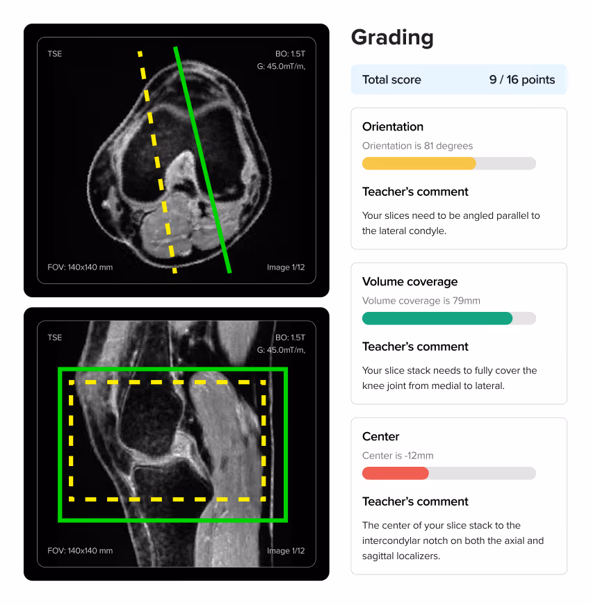
With auto-grading, you perform a reference scan on the simulator yourself. That image becomes the benchmark. Corsmed then automatically compares each student's submission against it and assigns points based on how close they got. A near-perfect result earns full marks. Students get instant feedback without you reviewing every scan individually.

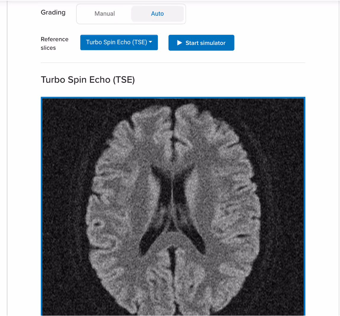
Example of a reference image on an auto-graded scan exercise:

Corsmed UI: a scan assignment block that uses auto-grading, teacher has uploaded an axial T2 TSE of a brain as reference image

Example of an auto-graded scan:

Screenshot of Corsmed's automated grading panel for a knee MRI exercise, showing a student's slice planning scored at 9 out of 16 points with instant feedback on orientation, volume coverage, and centering.

When using auto-grading, you can also specify how the platform should assess students' scans:

  1. What should be assessed:Parameters, slice planning, or both
  2. How strict or lenient the grading should be:The more lenient, the more a student's image can deviate from the reference while still earning a full score.

Corsmed's auto-grading functionality:

Screenshot of Corsmed's auto-grading functionality, showing two button groups. The first button group lets you choose what should be assessed (parameters, slice planning or both), and the second button group lets you choose how strict or lenient the grading should be (Low, Normal or High).

With manual grading, you review each submission yourself and write feedback directly on it, noting exactly what the student got right and what to correct. Students see your annotations alongside their scan.

How long does setup and onboarding take?

Most programs are up and running within a week. Corsmed is cloud-based, so there's no hardware to install and no IT configuration required. Users access everything through a web browser.

We provide onboarding sessions to help you structure your training program, drawing on what's worked at other hospitals and colleges. You're not building from scratch.

NHS UCLH found that a 1-hour introduction is enough for radiographers to begin practicing independently after that.

Is it difficult to set up and integrate into our training program?

No, setup is extremely simple. Corsmed works on any computer with an internet connection. There's no software to install, no special hardware to buy, and no IT configuration required. Users just log in and access it through a web browser.

NHS UCLH found that a 1-hour introduction is enough for radiographers to begin practicing independently.

"It's straightforward: press 'Scan' and it scans. Click 'Add protocol' and it adds a protocol.

A 1-hour introduction enables our learners to explore independently. There are good video guides within the 'Help' page too."

Noyal Mathew

Noyal Mathew

MRI Practice Educator, NHS UCLH

Even non-technical users learn to use Corsmed very quickly.

"One of our students wasn't very tech-savvy. She needed IT support just to upload her coursework. But she mastered Corsmed, which shows how user-friendly the simulator is."

Dr. Mahmud Khokhar

Dr. Mahmud Khokhar

MRI Program Director, CNI College

Will our students and technologists actually use it, or will it sit unused?

Yes, when integrated correctly. Usage depends heavily on how the simulator is introduced into your program.

NHS UCLH found that structured group sessions work best: learners practice together with an educator present for real-time feedback. When used in formal training sessions with clear exercises and learning objectives, engagement is strong. We share best practices from successful programs to help you get that right from the start.

How do we know if users are actually progressing?

The analytics dashboard gives you a live view of your whole cohort. Module completion, time spent on the simulator, scan count, and grades for every student are visible at a glance.

You can drill into any individual to see exactly where they are in the curriculum and which exercises they've completed. In a self-paced program, this lets you catch who is falling behind before it becomes a problem.

What if we run into technical issues or need help?

You get full support. Every account includes direct access to our support team and built-in help resources, including workflow videos and guides.

We also offer onboarding sessions to help you structure your training program around the simulator, based on what has worked for other hospitals and colleges. You are never on your own.