
Get FREE Weekly MRI Content


If you're an MRI educator, you've heard this same anxiety from your students.
They study the theory. They understand the equations. But when it's time to enter a real clinic and scan actual patients, they freeze.
The problem isn't the students. It's the training gap.
Students need hundreds of hours adjusting parameters, seeing how images change, and building the muscle memory that only comes from repetition. But real scanners are unavailable for this kind of practice.
Hospitals can't block scanner time for training when each hour represents $500–$2,000 in lost patient revenue. Even when you secure clinical placements, students mostly watch from the sidelines.
This creates an impossible situation: Students need practice to feel confident, but they can't get practice until they're already confident enough to work with real patients.
MRI simulation software is the solution to this theory-practice gap.
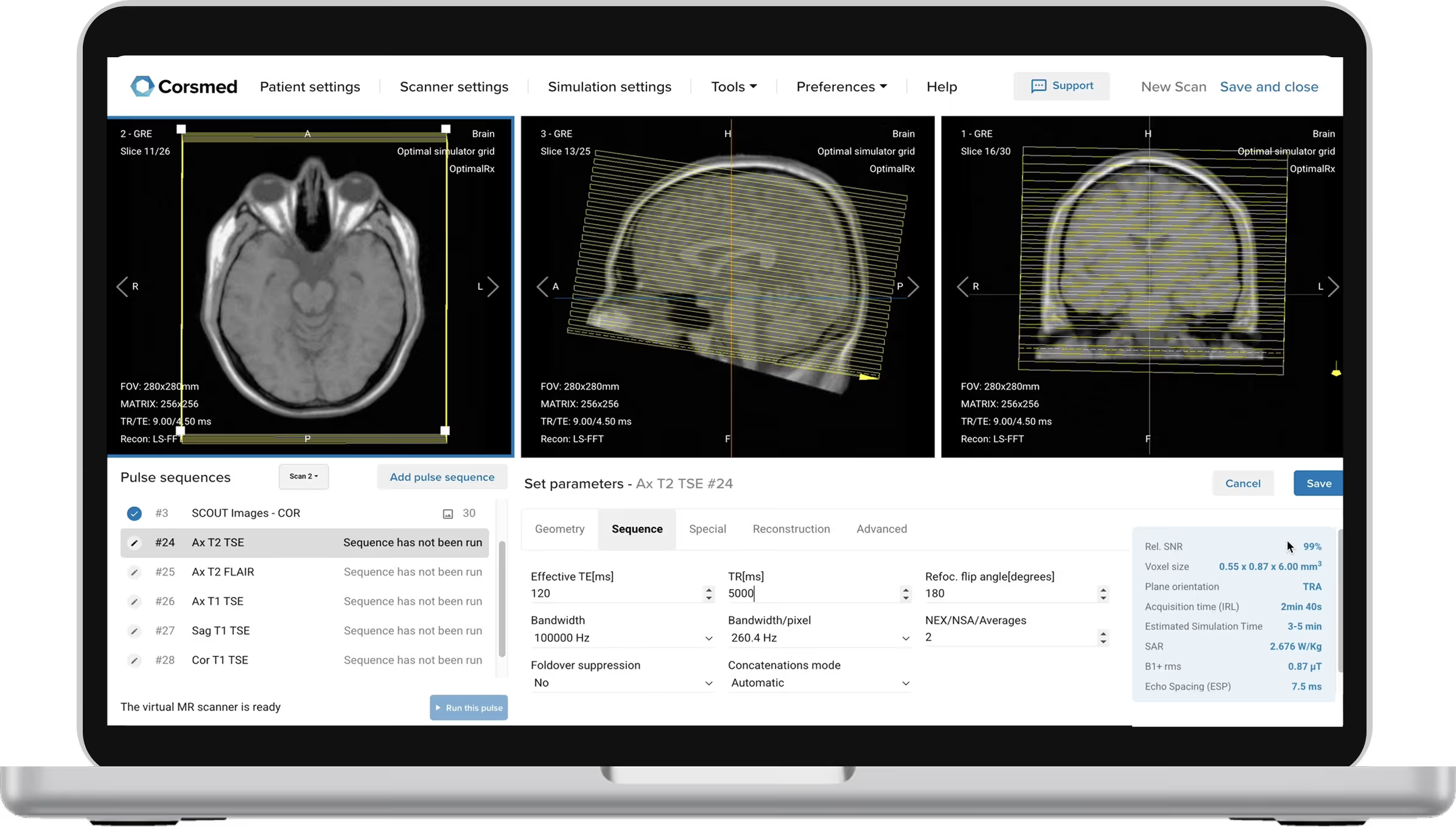
MRI simulators are digital tools that recreate the experience of operating a real scanner on your computer.
Students practice scanning anytime, anywhere. They adjust TR, TE, slice angles, and other parameters, then see exactly what image those settings would produce.
The best simulators create images from scratch using the same physics as real scanners. Change TE by 1ms, and the image updates exactly as it would in a real clinic.
MRI simulators improve training in five key ways:
This is the primary benefit of MRI simulators: they bridge the gap between classroom learning and clinical practice.
Teachers demonstrate concepts instead of just explaining them. And students change parameters and see instantly how the image changes.
Real scanner training requires being on-site during limited hospital hours. Simulators are available 24/7 from any device, letting students practice as much as they need.
Most students only see routine scans during training. Complex cases like cardiac MRI or MRCP might appear 3-4 times a year.
Real scanners take 3-5 minutes per sequence, while most simulators produce images in seconds.
The difference in practice volume is clear:
This 3-10X increase in practice volume cuts training time nearly in half.
You can't experiment on real patients, but learning requires making mistakes.
Simulators let students push parameters to extremes, create artifacts on purpose, and learn from errors without any consequences.
For simulation to prepare students for real clinical work, three factors are essential:
When you change parameters, does the image update exactly as it would on a real scanner?
Many simulators let you adjust settings, but the image stays the same. That's like a car with pedals but no engine.
Can you adjust field strength, coils, and work with multiple pulse sequences? Can you practice on brain, cardiac, MSK, and abdomen?
Perfect accuracy with limited coverage teaches very little. You need comprehensive clinical scenarios.
Does it work like a real scanner? Can you plan slices with your mouse and use an industry-standard interface?
If the workflow differs from clinical practice, students will struggle to operate real scanners.
All these three factors are necessary for simulated practice to transfer to real clinical competence on the scanner.
Corsmed is the only simulator meeting all requirements for clinical training as of 2026.
Here is how Corsmed fulfills the three key factors:
100% graduation rate. 100% employment rate.
Students hired before graduation, often with signing bonuses.
MRI enrollment doubled after implementing Corsmed.
Students are "10 times" more skillful when they graduate.
The American Registry of Magnetic Resonance Imaging Technologists (ARMRIT) has approved Corsmed training as part of required clinical hours.
Students can complete up to 250 of their required 1,000 hours using the simulator.
The institutions adopting clinical-grade simulation now will graduate the most skilled technologists.
Their students will be ready to address the workforce shortage and provide excellent patient care from Day One.
Get in touch with us today, and discover how Corsmed can take your MRI training to the next level.
More Success Stories:
Content
Content
Content
Content